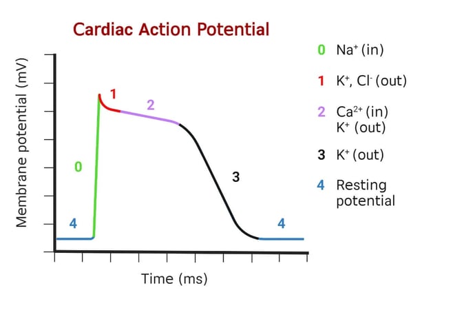

The action potential of the heart refers to the electrical impulses that trigger heart muscle contraction, ensuring coordinated heartbeats. The heart’s ability to contract in a rhythmic and controlled manner is due to the electrical activity that propagates through the heart muscle. This electrical activity is primarily generated and propagated by specialized cardiac cells, particularly within the sinoatrial (SA) node, atrioventricular (AV) node, bundle of His, bundle branches, and Purkinje fibers.
Phases of the Heart's Action Potential:
The action potential of the heart muscle cells (myocytes) is divided into five main phases, labeled 0-4. These phases reflect the flow of ions in and out of the heart muscle cells, generating and propagating the electrical signal that leads to contraction.
1. Phase 0: Depolarization (Rapid Upstroke)
Ion Movement: This phase begins when the action potential arrives at the myocyte and causes the fast sodium (Na⁺) channels to open, allowing sodium ions to rush into the cell.
Effect: The rapid influx of sodium ions causes the cell to become more positive inside, leading to a rapid depolarization of the membrane (from -90 mV to +30 mV).
2. Phase 1: Initial Repolarization
Ion Movement: During this phase, the sodium channels close, and potassium (K⁺) channels open, allowing potassium ions to move out of the cell.
Effect: There is a brief partial repolarization, where the membrane potential begins to drop slightly (but not all the way back to its resting potential).
3. Phase 2: Plateau Phase
Ion Movement: In this phase, the calcium (Ca²⁺) channels open, allowing calcium ions to enter the cell, while potassium continues to leave the cell. This balance between the influx of calcium and the efflux of potassium causes the membrane potential to stabilize at a relatively positive level (about 0 mV).
Effect: This plateau phase is crucial because it extends the duration of the action potential, leading to a longer contraction. The influx of calcium triggers the release of more calcium from the sarcoplasmic reticulum, which is essential for muscle contraction (the process of excitation-contraction coupling).
4. Phase 3: Repolarization
Ion Movement: As the calcium channels close, potassium channels remain open, allowing potassium to move out of the cell. The cell becomes more negative inside, leading to repolarization.
Effect: The membrane potential returns toward its resting state (around -90 mV). This phase marks the return of the cell to its resting electrical state and prepares it for the next action potential.
5. Phase 4: Resting Membrane Potential
Ion Movement: During this phase, the cell maintains a resting membrane potential of about -90 mV, primarily due to the sodium-potassium pump (Na⁺/K⁺ ATPase), which actively pumps sodium out of the cell and potassium back in.
Effect: The cell is at rest, ready to be depolarized again by a new action potential.
Key Points
Phase 0 (Depolarization): Rapid Na⁺ influx → membrane potential increases.
Phase 1 (Initial Repolarization): Na⁺ channels close, K⁺ efflux causes slight repolarization.
Phase 2 (Plateau): Ca²⁺ influx balances K⁺ efflux → plateau phase.
Phase 3 (Repolarization): K⁺ efflux dominates, membrane potential returns to rest.
Phase 4 (Resting Potential): Stable resting potential maintained by Na⁺/K⁺ ATPase pump.
Anatomy Useful Links
