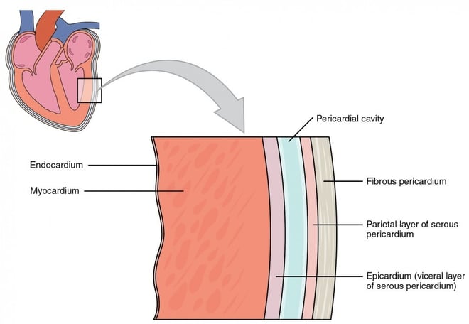

The heart is made up of three distinct layers, each with its own specific function to ensure proper heart function and circulation. These layers are:
1. Endocardium
Location: The innermost layer of the heart, lining the heart chambers, valves, and blood vessels that enter and exit the heart.
Structure: The endocardium is composed of a smooth layer of epithelial cells called endothelial cells, which is supported by a thin layer of connective tissue.
Function:
It helps prevent blood clotting by providing a smooth, frictionless surface.
It forms the lining of the heart valves, ensuring proper valve function and reducing the risk of blood flow disturbances.
2. Myocardium
Location: The middle and thickest layer of the heart, located between the endocardium and the epicardium.
Structure: The myocardium consists of cardiac muscle tissue, which is unique for its ability to contract rhythmically and efficiently. It is the muscular layer responsible for the heart’s pumping action.
Function:
The myocardium is responsible for the contraction of the heart, which pumps blood throughout the body.
It has a high density of mitochondria to meet the energy demands of continuous contraction.
The thickness of the myocardium varies: it is thicker in the left ventricle (as it pumps blood to the entire body) than in the right ventricle (which only pumps blood to the lungs).
3. Epicardium
Location: The outermost layer of the heart, also known as the visceral pericardium.
Structure: The epicardium consists of a layer of epithelial cells and a thin layer of connective tissue. It also contains blood vessels, nerves, and fat that help protect the heart.
Function:
The epicardium serves as a protective layer for the heart.
It helps anchor the heart to the surrounding structures, such as the diaphragm and blood vessels.
It produces a small amount of fluid that lubricates the heart's surface, reducing friction as the heart beats within the pericardial sac.
4.Pericardium (Not one of the heart layers, but important):
While the pericardium is not a heart layer itself, it plays a critical protective role. It is a double-layered membrane surrounding the heart, consisting of:
Fibrous pericardium: The tough outer layer that prevents overstretching of the heart.
Serous pericardium: A thinner, inner layer that is further divided into the parietal and visceral pericardium (the visceral layer is also the epicardium of the heart).
Summary of the Layers:
Endocardium – Innermost, smooth lining of heart chambers and valves.
Myocardium – Middle muscular layer responsible for heart contraction.
Epicardium – Outermost layer, protective and lubricative.
Together, these layers work to ensure that the heart can pump blood effectively while being protected from damage and friction during each heartbeat.
Key points:
Epicardium: Outermost Layer, thin layer of connective tissue. Protect the heart.
Myocardium: Middle Layer, thick layer of cardia muscle(cardiomyocytes). Pumps blood throughout the body.
Endocardium: Innermost layer, thin layer of endothelial cells. Lines heart chambers, valves and blood vessels.
pericardium: Double-Layered sac surrounding the heart. Protects and anchors the heart.
Anatomy Useful Links
