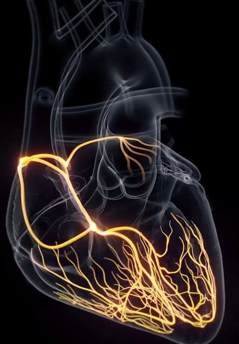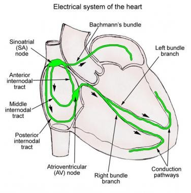

The conduction system of the heart is a network of specialized cells responsible for generating and conducting electrical impulses that control the heart's rhythm and coordinate its contractions. The system ensures that the heart beats in a synchronized manner, allowing it to pump blood efficiently. Here’s a breakdown of the key components of the heart’s conduction system:
1. Sinoatrial (SA) Node
Location: Right atrium, near the opening of the superior vena cava.
Function: Known as the "natural pacemaker" of the heart, the SA node initiates electrical impulses. These impulses spread across both atria, causing them to contract (atrial systole).
Rate: The SA node generates impulses at a rate of about 60-100 beats per minute in a resting adult, though this can be influenced by factors like the autonomic nervous system.
2. Atrioventricular (AV) Node
Location: At the junction of the atria and ventricles, near the tricuspid valve.
Function: The AV node acts as a gatekeeper, receiving the electrical impulse from the SA node and briefly delaying it. This delay allows the atria to finish contracting and the ventricles to fill with blood before they contract.
Rate: If the SA node fails, the AV node can take over as the pacemaker, but at a slower rate, typically around 40-60 beats per minute.
3. Bundle of His
Location: Located in the interventricular septum, just below the AV node.
Function: The Bundle of His is a pathway that conducts the electrical impulse from the AV node to the right and left bundle branches.
Rate: It conducts the impulse rapidly to ensure that the ventricles contract in a coordinated manner.
4. Right and Left Bundle Branches
Location: These branches extend down the interventricular septum, carrying the impulse to the right and left ventricles, respectively.
Function: They transmit the electrical signals to the ventricles, ensuring that both ventricles contract simultaneously.
Rate: These branches conduct the impulse quickly to the Purkinje fibers.
5. Purkinje Fibers
Location: Located in the walls of the ventricles.
Function: The Purkinje fibers distribute the electrical impulse throughout the ventricles, leading to coordinated ventricular contraction (ventricular systole).
Rate: The Purkinje fibers themselves can act as a pacemaker at a very slow rate (20-40 beats per minute) if necessary.
Electrical Conduction Pathway:
Your heart’s conduction system is like the electrical wiring of a building. It controls the rhythm and pace of your heartbeat. Signals start at the top of your heart and move down to the bottom. Your conduction system includes:
SA Node → Atria (contraction) → AV Node (delay) → Bundle of His → Right and Left Bundle Branches → Purkinje Fibers → Ventricles (contraction).
Sinoatrial (SA) node: Sends the signals that make your heartbeat.
Atrioventricular (AV) node: Carries electrical signals from your heart’s upper chambers to its lower ones.
Left bundle branch: Sends electric impulses to your left ventricle.
Right bundle branch: Sends electric impulses to your right ventricle.
Bundle of His: Sends impulses from your AV node to the Purkinje fibers.
Purkinje fibers: Make your heart ventricles contract and pump out blood.
Role of the Conduction System:
Rhythmic heartbeat: Ensures that the heart beats regularly and efficiently.
Coordinated contraction: Controls the timing of contractions between the atria and ventricles.
Autonomic regulation: The autonomic nervous system can modulate the heart rate by influencing the conduction system. The sympathetic nervous system speeds up the heart rate, while the parasympathetic system slows it down.
The proper functioning of the heart's conduction system is crucial for normal cardiac function, and disruptions in this system can lead to arrhythmias or other heart conditions.
Key Points
SA Node is the heart's natural pacemaker, setting the pace of the heart.
The AV Node provides a brief delay, ensuring the atria have time to contract before the ventricles contract.
The Bundle of His and bundle branches direct the impulse down to the ventricles.
Purkinje fibers spread the impulse throughout the ventricles to initiate ventricular contraction.
Anatomy Useful Links
Conduction System of the Heart


