Function
What is the function of the heart?
Your heart’s main function is to move blood throughout your body. Blood brings oxygen and nutrients to your cells. It also takes away carbon dioxide and other waste so other organs can dispose of them.
Your heart also:
Controls the rhythm and speed of your heart rate.
Maintains your blood pressure.
Your heart works with these body systems to control your heart rate and other body functions:
Nervous system: Your nervous system helps control your heart rate. It sends signals that tell your heart to beat slower during rest and faster during stress.
Endocrine system: Your endocrine system sends out hormones. These hormones tell your blood vessels to constrict or relax, which affects your blood pressure. Hormones from your thyroid gland can also tell your heart to beat faster or slower.
The human heart is a vital organ responsible for pumping blood throughout the body. It functions as part of the circulatory system, ensuring that oxygen, nutrients, and waste products are transported to and from cells, tissues, and organs. It beats around 100,000 times per day, making it one of the hardest working muscles in the body.
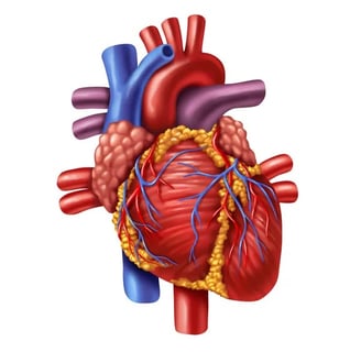
The Heart is a fist-sized organ in the thoracic cavity pumping blood and supplying and nutrients. It has four chambers, four valves and blood vessels, with a heartbeat of 70-80bpm. Essential for life, it regulates blood pressure and maintains cardiac output. Here's an overview of its anatomy:
Structure of the Heart
The heart is divided into four chambers:
Right Atrium: Receives deoxygenated blood from the body through the superior and inferior vena cava.
Right Ventricle: Pumps deoxygenated blood to the lungs via the pulmonary artery for oxygenation.
Left Atrium: Receives oxygenated blood from the lungs through the pulmonary veins.
Left Ventricle: Pumps oxygenated blood to the entire body through the aorta.
Valves
The heart has four main valves that prevent backflow of blood:
Tricuspid Valve: Between the right atrium and right ventricle.
Pulmonary Valve: Between the right ventricle and the pulmonary artery.
Mitral Valve (Bicuspid Valve): Between the left atrium and left ventricle.
Aortic Valve: Between the left ventricle and the aorta.
Blood Flow Pathway
Deoxygenated Blood enters through the superior and inferior vena cava into the right atrium.
It then passes through the tricuspid valve to the right ventricle.
The right ventricle pumps the blood through the pulmonary valve into the pulmonary artery, which transports the blood to the lungs for oxygenation.
Oxygenated blood returns from the lungs through the pulmonary veins into the left atrium.
The blood moves through the mitral valve into the left ventricle.
The left ventricle pumps the oxygen-rich blood through the aortic valve into the aorta, which distributes the blood to the rest of the body.
Heart Wall Layers
Epicardium: The outer layer, which also serves as the protective membrane.
Myocardium: The thick middle layer made of cardiac muscle responsible for contraction.
Endocardium: The inner lining of the heart, which is smooth and prevents blood clotting.
Functions of the Heart
Pumping Blood: Circulates blood to supply oxygen and nutrients to tissues and remove carbon dioxide and waste.
Heartbeat: Regular contractions controlled by electrical impulses ensure continuous circulation.
Regulation of Blood Pressure: The heart adjusts the force of contractions to maintain blood pressure.
Cardiac Cycle
The cardiac cycle is the sequence of events that occurs when the heart beats. It consists of two main phases:
Systole: The contraction phase, where the heart pumps blood out of the chambers.
In ventricular systole, the ventricles contract, pushing blood into the pulmonary artery (from the right ventricle) and the aorta (from the left ventricle).
During atrial systole, the atria contract, filling the ventricles with blood.
Diastole: The relaxation phase, where the heart chambers fill with blood in preparation for the next contraction.
In ventricular diastole, the ventricles relax and fill with blood from the atria.
In atrial diastole, the atria relax, awaiting the next impulse to contract.
Heart Sounds
The characteristic "lub-dub" sounds of the heart are produced by the closing of heart valves:
Lub (S1): The sound of the mitral and tricuspid valves closing as the ventricles begin to contract.
Dub (S2): The sound of the aortic and pulmonary valves closing as the ventricles finish contraction.
Blood Vessels
The heart is connected to a vast network of blood vessels that circulate blood throughout the body:
Arteries: Carry oxygenated blood away from the heart.
The aorta is the largest artery, delivering oxygen-rich blood to the body.
The pulmonary artery carries deoxygenated blood to the lungs for oxygenation.
Veins: Carry deoxygenated blood back to the heart.
The superior and inferior vena cavae return deoxygenated blood from the body to the right atrium.
The pulmonary veins bring oxygenated blood from the lungs to the left atrium.
Capillaries: Tiny blood vessels where oxygen, nutrients, and waste products are exchanged between blood and tissues.
Heart Rate and Cardiac Output
Heart Rate (HR): The number of times the heart beats per minute. A normal resting heart rate for adults is typically between 60-100 beats per minute.
Cardiac Output (CO): The volume of blood the heart pumps per minute. It is the product of stroke volume (SV), which is the amount of blood pumped per beat, and heart rate (HR). The formula is:
CO=SV×HRCO = SV \times HRCO=SV×HR
Anatomy Of Human Heart
Our team
Our strength lies in our individuality. Set up by Esther Bryce, the team strives to bring in the best talent in various fields, from architecture to interior design and sales.
Esther Bryce
Founder / Interior designer
Lianne Wilson
Broker
Jaden Smith
Architect
Jessica Kim
Photographer